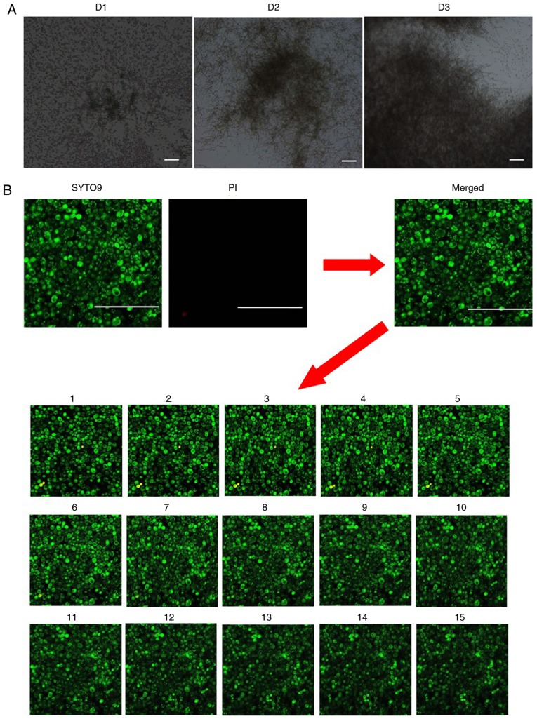
Figure 3.
Effects of blue light on C. albicans inside biofilms. (A) Optical microscope images of C. albicans biofilms grown in the wells of plates. D1 represents the early phase, D2 represents the intermediate phase and D3 maturation phase of C. albicans biofilm formation. (Magnification, ×100; scale bars, 100 µm). (B) Distribution of the dead/viable C. albicans in the biofilm of the control group as observed by confocal laser scanning microscopy (Magnification, ×400; scale bars, 50 µm). Green staining indicates live C. albicans, and red staining indicates dead C. albicans. Images 1–15 represent scanning images from the deepest layer of the biofilm to the superficial layers of the biofilm. C. albicans, Candida albicans; PI, propidium iodide.

