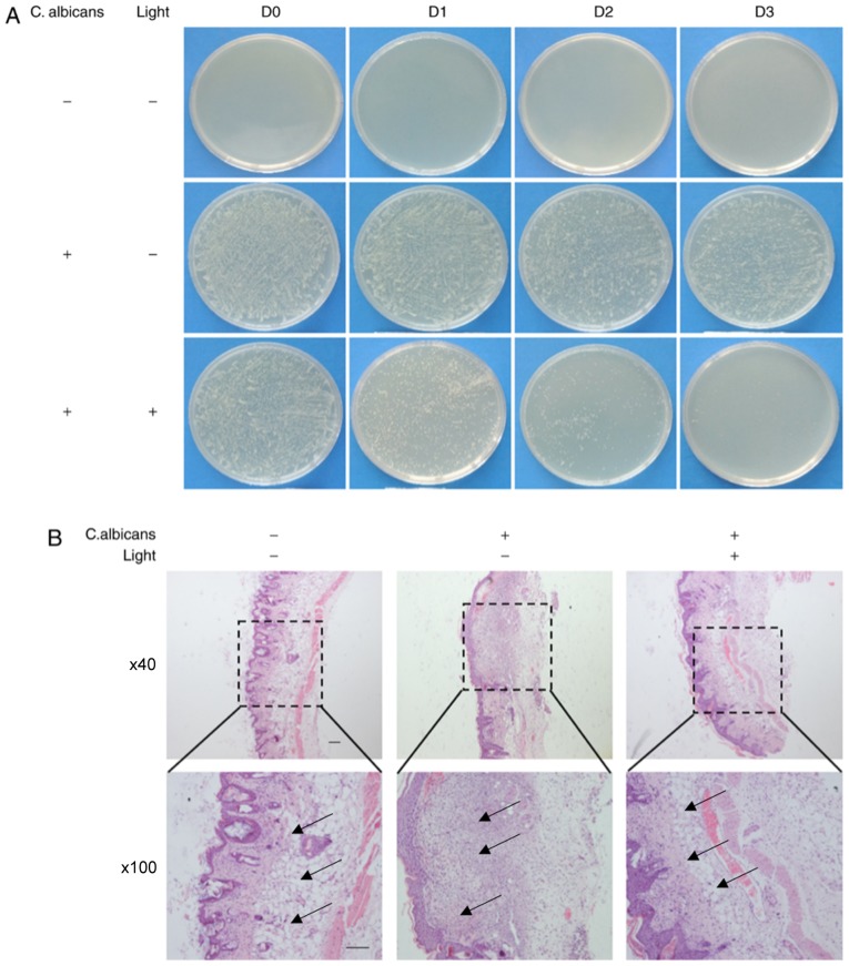
Figure 5.
Investigation into the effects of BL on established models of mice wound healing following inoculation with C. albicans in vivo. (A) Images of spread plates of the mice wound secreta obtained from the control group (C. albicans -, Light -), the untreated C. albicans group (C. albicans +, Light-) and the C. albicans group treated with 240 J/cm2 BL (C. albicans +, Light +) at 0, 1, 2 and 3 day time intervals. (B) Hematoxylin and eosin staining of wound tissues at day 7. Black arrow: Infiltration of inflammatory cells. (Scale bars, 100 µm). BL, blue light; C. albicans, Candida albicans.

