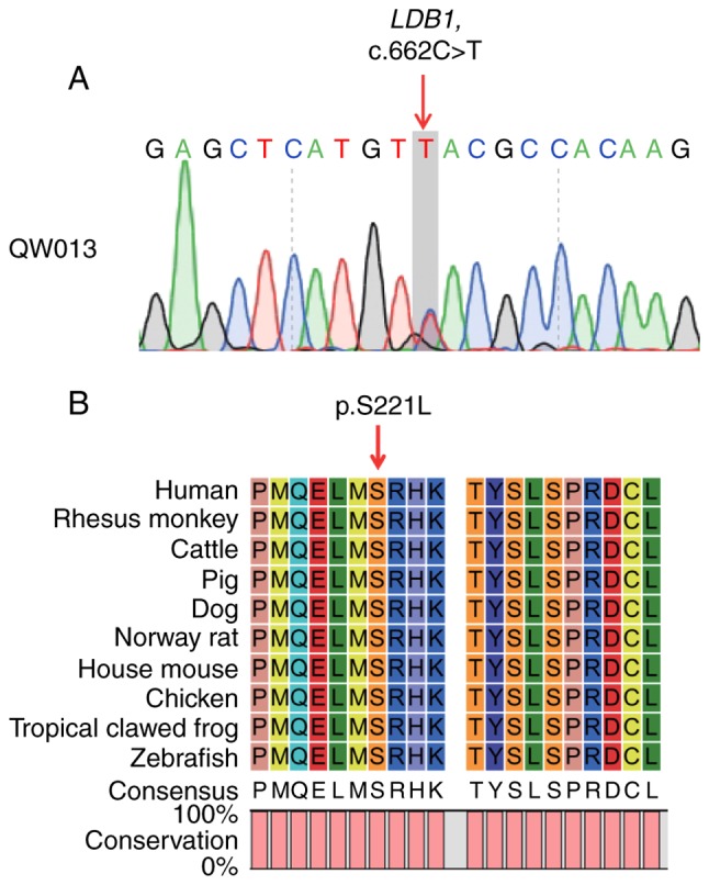
Figure 2.

Analysis of the LDB1 variant. (A) Sanger sequencing validated the heterozygous c.662C>T variant in the LDB1 gene. The red arrow indicates the mutation site. (B) Amino acid sequence alignment of LDB1 in different species. The red arrow indicates the mutational amino acid. Serine at position 211 was 100% conserved (full red columns) in all species. LDB1, LIM domain-binding protein 1.
