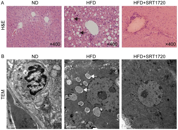
Figure 1.
H&E staining and TEM images of liver tissues. (A) Histopathologic changes of liver tissues in each group (magnification, ×400). There was no obvious liver injury in control group. Extensive ballooning degeneration of hepatocytes and infiltration of inflammatory cells were displayed in HFD group. Fewer ballooning degeneration of hepatocytes and infiltration of inflammatory cells were observed in HFD+SRT1720 group. (B) The ultrastructure of mice liver from HFD+SRT1720 group was less severe than in HFD group. Arrows, steatosis and lipid droplet. H&E, hematoxylin and eosin; HFD, high-fat diet; ND, normal diet.

