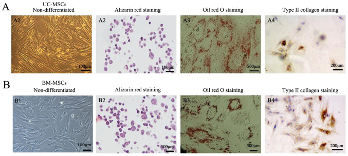
Figure 3.
Detection of the differentiation potential of (A) UC-MSCs and (B) BM-MSCs. (A1) UC-MSCs and (B1) BM-MSCs were investigated for their differentiation capacity. (A2 and B2) Osteogenic differentiation was examined by alizarin red staining. Scale bars, 1-µm. (A3 and B3) Adipogenic differentiation was examined by Oil Red O staining. Scale bars, 500 µm. (A4 and B4) Cells were differentiated into chondrogenic cells and immunohistochemical stained positive for type II collagen. Scale bars, 200 µm. UC-MSCs, umbilical cord mesenchymal stem cells; BM-MSCs, bone marrow derived mesenchymal stem cells.

