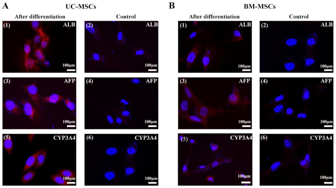
Figure 5.
Immunofluorescent analysis of hepatocyte-specific proteins. (A) UC-MSCs and (B) BM-MSCs were examined for their expression of (A1 and B1) ALB, (A3 and B3) AFP, and (A5 and B5) CYP3A4 following hepatic differentiation for 4 weeks. (A2, 4 and 6, and B2, 4 and 6) Cells cultured in the growth medium were as negative controls. Scale bars, 100 µm. UC-MSCs, umbilical cord mesenchymal stem cells; BM-MSCs, bone marrow derived mesenchymal stem cells; ALB, albumin; AFP, α-fetoprotein; CYP3A4, cytochrome P450 3A4.

