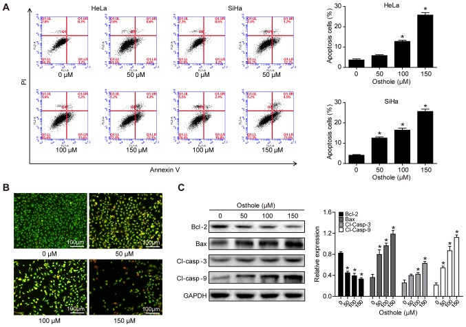
Figure 2.
Osthole induces cervical cancer cell apoptosis. (A) HeLa and SiHa cells were grown and treated with osthole (0, 50, 100 or 150 µM) for 24 h and subjected to the apoptosis assay. (B) Tumor cell AO/EB fluorescence staining. HeLa cells were grown and treated with osthole (0, 50, 100 or 150 µM) for 24 h and subjected to staining. (C) Western blot analysis. Tumor cells were grown and treated with or without osthole (0, 40, 80, 120, 160, 200 or 240 µM) for 24 h and then subjected to western blot analysis of Bcl-2, Bax, and cleaved caspase-3 and −9 proteins. *P<0.05 compared to the control group. Bcl-2, B-cell lymphoma 2; Bax, Bcl-2-associated X protein; Cl, cleaved.

