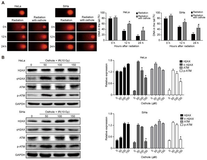
Figure 5.
Osthole induces cervical cancer cell DNA damage induced by radiation. (A) Comet assay. HeLa and SiHa cells were grown and treated with 50 µM osthole for 24 h and then subjected to 6 Gy irradiation and then subjected to the Comet assay. A total of 50 cells were randomly quantified from the images and the percentage of cell tail length was calculated. Data are summarized as the mean ± standard deviation. *P<0.05 compared to the control. (B) Western blot analysis. HeLa cells were grown and treated with osthole (50,100 or 150 µM) for 24 h and exposed to 10 Gy radiation and then subjected to western blot analysis for the detection of p-ATM, ATM, γH2AX and H2AX. ATM, ataxia telangiectasia mutated; p-, phosphorylated; IR, irradiation.

