Figure 6.
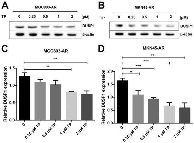
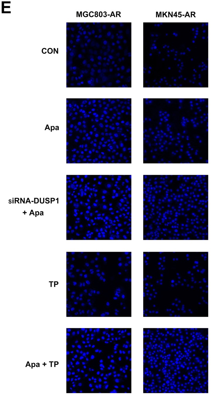
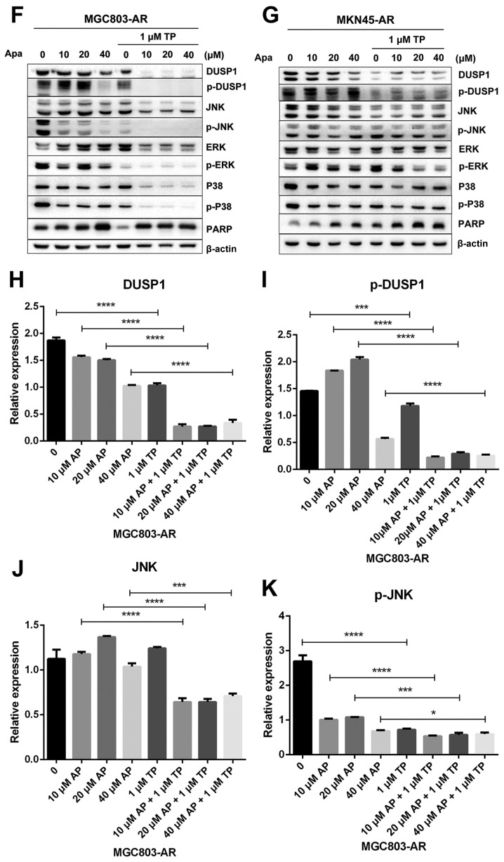
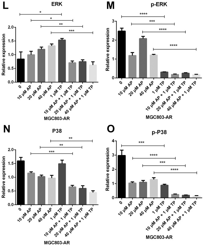
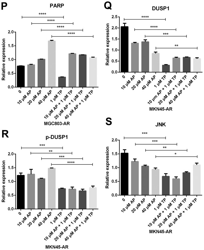
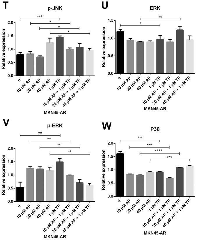
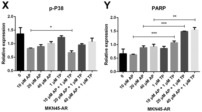
Triptolide combined with Apa overcomes Apa resistance by inhibiting MAPK signaling and inducing apoptosis. (A) MGC803-AR and (B) MKN45-AR cells were treated with a concentration gradient of triptolide, and expression of DUSP1 was evaluated by western blot analysis using β-actin as a loading control. Histograms show the relative quantitative expression in (C) MGC803-AR and (D) MKN45-AR cells. Data are presented as the mean ± standard deviation (n=3; Student's t-test; *P<0.05, **P<0.01, ***P<0.001, ****P<0.0001). (E) MGC803-AR and MKN45-AR cells were treated with 10 µM Apa and cells with DUSP1 knockdown were treated with 10 µM Apa or 1 µM triptolide alone or 10 µM Apa + 1 µM triptolide for 6 h. Cells were stained with Hoechst 33342 and images were captured using an Olympus BH-2 fluorescence microscope (magnification, ×40). (F) MGC803-AR and (G) MKN45-AR cells were treated with Apa or Apa + 1 µM triptolide for 24 h at different Apa concentrations. Total cell lysates were prepared and analyzed by western blot analysis using antibodies directed against MAPK signaling molecules (H) DUSP1, (I) p-DUSP1, (J) JNK and (K) p-JNK. (L) ERK, (M) p-ERK, (N) P38, (O) p-P38, (P) PARP in MGC803-AR cells; and (Q) DUSP1, (R) p-DUSP1, (S) JNK. and (T) p-JNK (U) ERK, (V) p-ERK, (W) P38, (X) p-P38, (Y) PARP in MKN45-AR cells. β-actin was a loading control. Data are presented as the mean ± standard deviation (n=3; Student's t-test; *P<0.05, **P<0.01, ***P<0.001, ****P<0.0001). Apa/AP, apatinib; AR, Apa resistant; MAPK, mitogen-activated protein kinase; p-, phosphorylated DUSP, dual-specificity phosphatase-1; JNK, c-Jun N-terminal kinase; ERK, extracellular signal-regulated kinase; p-, phosphorylated; PARP, poly(ADP-ribose) polymerase.

