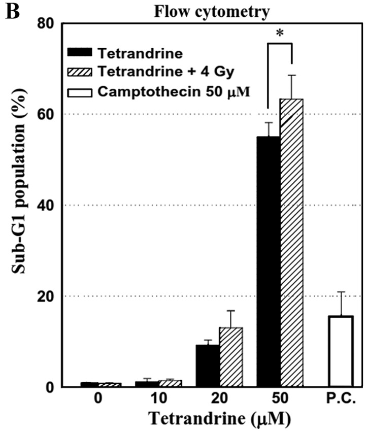
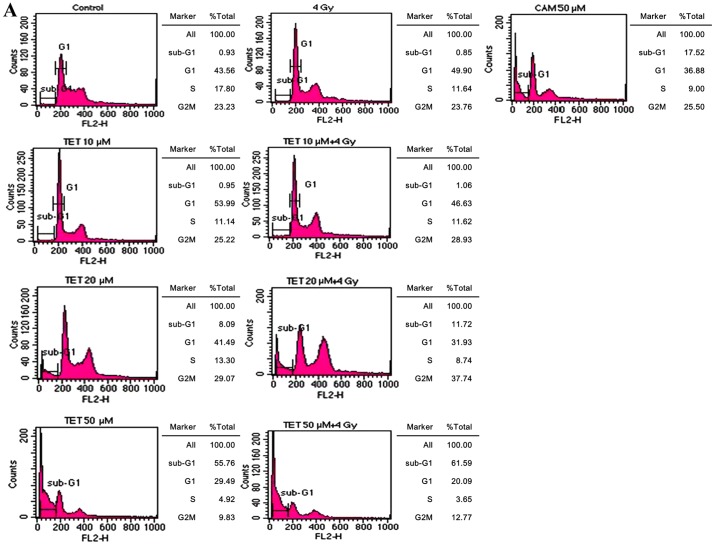
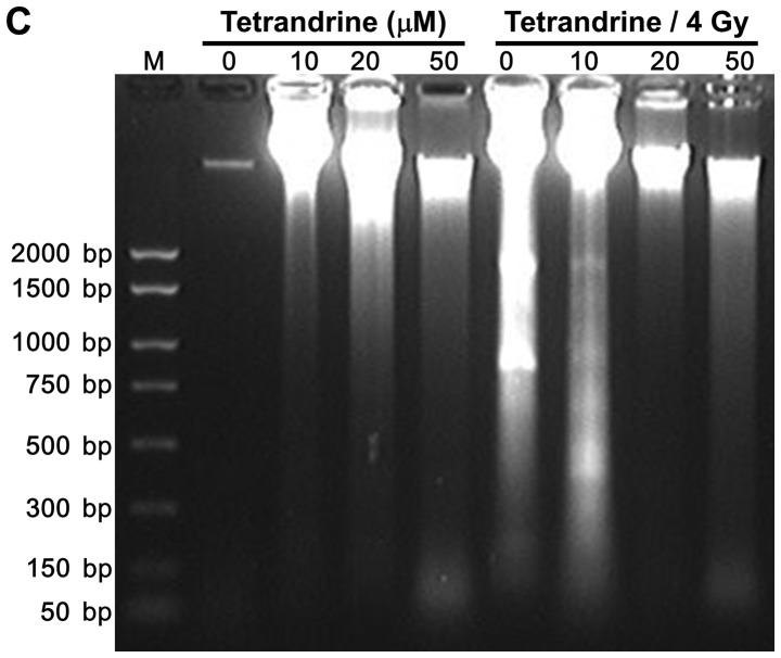
Figure 2.

Cell cycle analysis of CT26/tk-luc cells treated with tetrandrine (TET) and/or radiation. Cells were treated with various concentrations of TET alone for 3 h or combined with 4 Gy of radiation. DNA content was evaluated by (A) propidium iodide staining and (B) analyzed with a FACSCalibur flow cytometer. Camptothecin (CAM; 50 µM) was used as the positive control (P.C.). Representative results are presented. Sub-G1 populations obtained from three independent experiments were quantified to identify the percentage of apoptotic cells. (C) Agarose gel electrophoresis of DNA extracted from CT26/tk-luc cells treated with various concentrations of TET alone or combined with 4 Gy of radiation. DNA fragmentation was more evident in the combination group. M, marker. The numbers on the y-axis stand for base pairs. *P<0.05.


