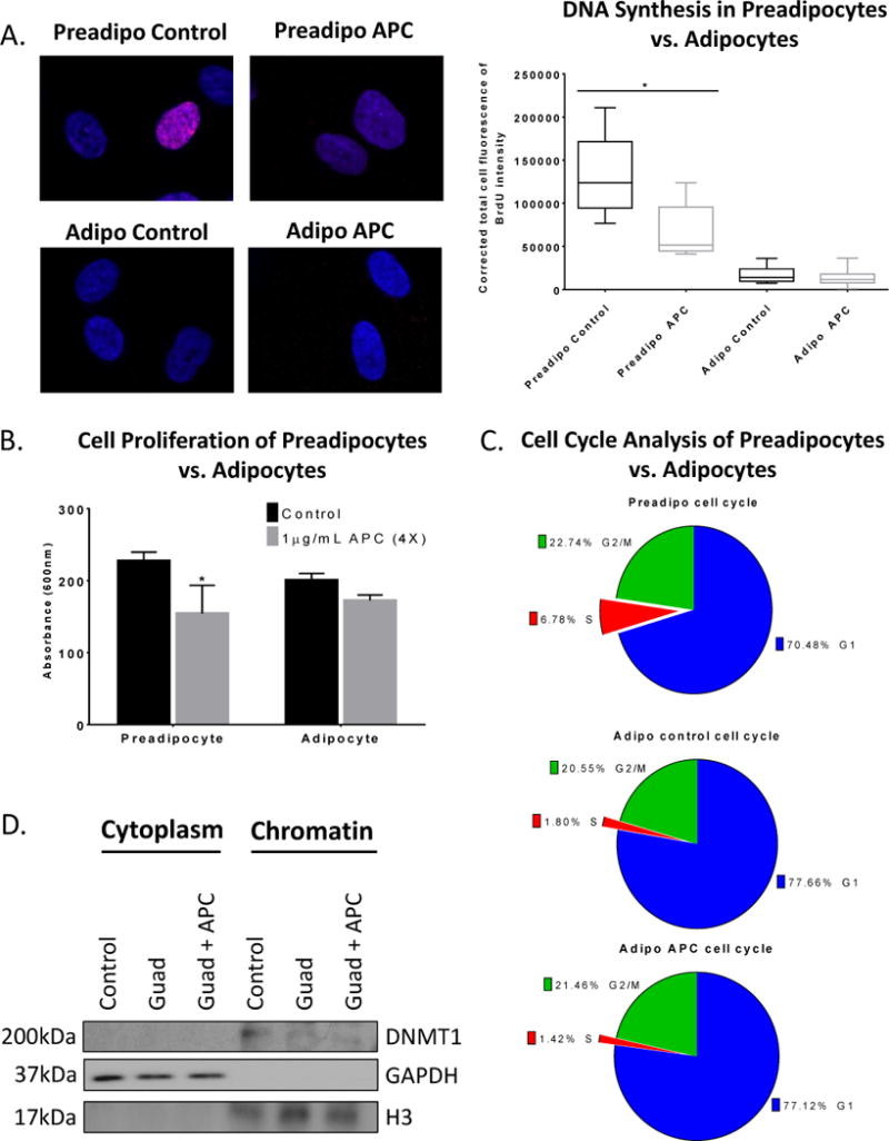
Figure 2. Guadecitabine degrades DNMT1 in the absence of DNA synthesis and cell division.

(A). BrdU immunofluorescence labeling (2h) was performed in preadipocytes and adipocytes treated with the DNA synthesis inhibitor aphidicolin (APC) (1μg/mL, daily 4X). (B) MTT cell proliferation assay was performed in preadipocytes and adipocytes treated with aphidicolin (APC) (1μg/mL, daily 4X). (C). Propidium iodide cell cycle flow cytometry analysis was performed on preadipocytes and adipocytes treated with APC (1μg/mL, daily 4X). (D). Subcellular fractionation was performed on adipocytes treated with either guadecitabine (100nM, daily 3X) alone or APC (1μg/mL, daily 4X) starting on day 1 and guadecitabine together with APC therein after for 3 days. Western blot was performed on cytoplasmic and chromatin fractions and probed for DNMT1, histone H3, and GAPDH. All experiments were performed in triplicate. (*P < 0.05).
