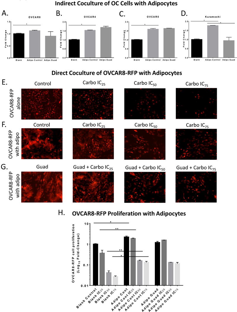
Figure 3. Adipocytes increase HGSOC proliferation.

(A). MTT proliferation assay of OVCAR8, (B) OVCAR4, (C) OVCAR5, (D) Kuramochi cells in the top of 0.45 micron Boyden chamber in indirect coculture with adipocytes treated with guadecitabine (100nM, daily 3X). (E). Images of OVCAR8-RFP cells plated directly in a 6-well plate and treated with carboplatin at IC25, IC50, and IC75 in serum-free media for 6d. (F). Images of OVCAR8-RFP cells directly cocultured with SGBS adipocytes (1:3 ratio) in serum-free media and treated with carboplatin IC25=10.67 μM, IC50=32 μM, and IC75=96μM for 6d. (G). Images of OVCAR8-RFP cells directly cocultured with SGBS adipocytes initially treated with guadecitabine (100nM, daily 3X) and removed, then OVCAR8-RFP cells and adipocytes were treated with carboplatin IC25, IC50, and IC75 for 6d. (H). Flow cytometry was performed to quantify number of OVCAR8-RFP when cocultured with SGBS adipocytes in comparison to control, guadecitabine treatment, and carboplatin treatment. All experiments were performed in triplicate. (*P < 0.05, **P < 0.01).
