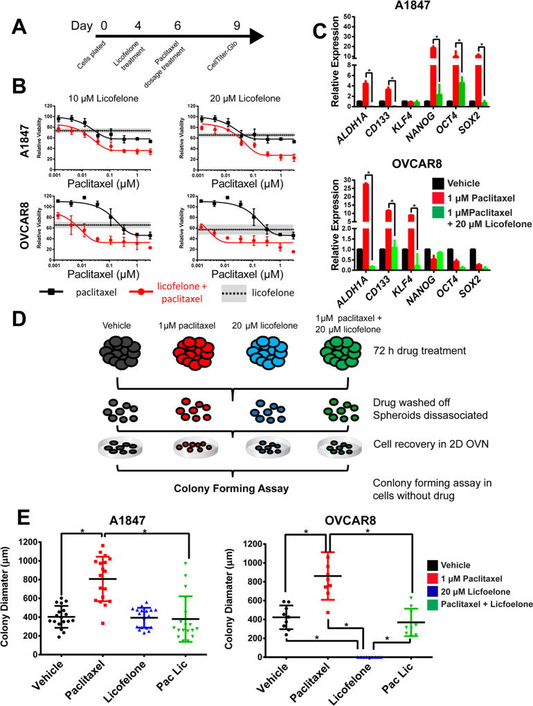
Figure 5. Licofelone synergistically enhances paclitaxel activity in combination while reversing the stem-like characteristics of ovarian MCTS.
(A) Representative time-line of drug combinations in A1847 and OVCAR8 spheroids. (B) Dosage response to paclitaxel alone (black line) is lower than paclitaxel following 72 h pretreatments with 10 µM licofelone (top) or 20 µM licofelone (bottom) (red lines). Licofelone alone treatments of 10 µM or 20 µM are represented by the horizontal dashed lines (mean) and grey boxes (SD). (C) Expression of cancer stem cell-related genes following 72 h treatment of vehicle (DMSO), 1 µM paclitaxel, or 1 µM paclitaxel and 20 µM licofelone in A1847 or OVCAR8 spheroids. (D) Graphical representation of secondary colony forming assays from MCTS. (E) Colony size (µm) was significantly increased in cells derived from previously paclitaxel treated MCTS and was decreased from cells derived from paclitaxel and licofelone treatments (n>10, *=p<0.05, two-way ANOVA). Colony formation assays were carried out in the absence of drug treatment. All data are represented as mean +/− SD, n=3.

