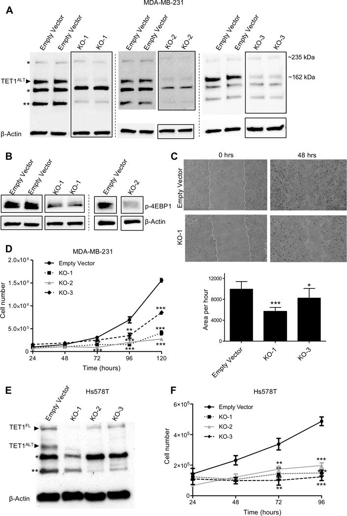
Figure 3. TET1 is essential for growth in TNBC.

(A) Western blot (in duplicate) of MDA-MB-231 empty vector and TET1 knockout single clones. β-actin used as a loading control. (* denotes putative non-specific band, ** denotes another potential truncated isoform of TET1) (B) Western blot of phospho-4EBP1 (Thr37/46) in MDA-MB-231 TET1 KO cells. KO-1 is in duplicate, KO-2 is single experiment. (C) Representative images of wound-healing assays in empty vector and TET1 KO-1 cells at 0 and 48 hours (top) and quantification (bottom), experiment performed in triplicate. (D) Cell proliferation assay for MDA-MB-231 empty vector, KO-1, KO-2 and KO-3. Cells were counted every 24 hours (in triplicate, each sample counted twice). X axis is time, Y axis is total cell number. (E) Western blot of empty vector and TET1 knockout single clones in Hs578T cells. (F) Cell proliferation assay for Hs578T empty vector, KO-1, KO-2 and KO-3. X axis is time, Y axis is total cell number.
