Figure 2.
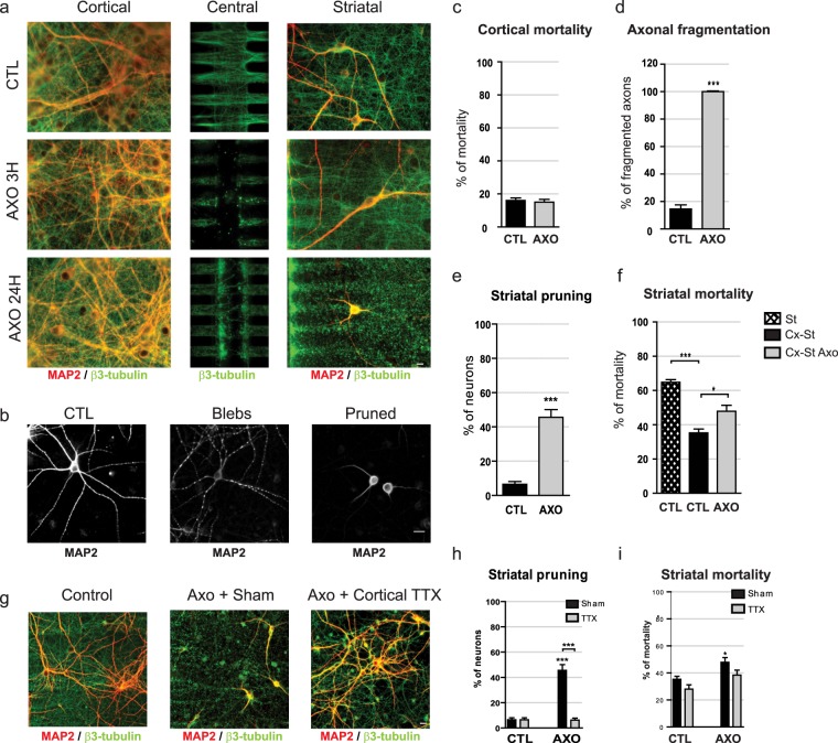
Cortical axotomy induces trans-synaptic degeneration in striatal neurons. (a) Cortical, central, and striatal chambers of normal (CTL) or axotomized (AXO3h, AXO24h) reconstructed cortico-striatal networks. Neurons were stained for MAP-2 (red), α-tubulin (green). Cortical neurons send axons efficiently through the micro-channels and the central chamber to reach, and connect with, striatal neurons. While cortical axons can be evidenced in the central chamber of CTL networks, 3 hours after axotomy the central chamber is void of axons, but axons inside the micro-channels and in the striatal chamber remain intact. Twenty-four hours after cortical axotomy all cortical axons disconnected from their soma are fragmented, and both the survival and the complexity of the dendritic arborization of remaining striatal neurons were altered (MAP-2, red) (Fig. 3a, lower panel). Scale bar 10 µM. (b) Striatal dendrites underwent dendritic beading called “blebs” followed by the regression of the dendritic arborization leading to a phenotype called “pruned”, with few and short dendrites compared to controls. Scale bar 10 µM. (c) Quantification of cortical mortality 24 hours post axotomy. (d) Quantification of cortical axonal fragmentation 24hours post-axotomy. (e) Quantification of striatal pruning, and striatal mortality (f) 24 hours post-axotomy. (g) Cortical somata and axons pretreated with TTX 5 min before and during the first 5 min after axotomy. After microchannel wash, cultures were further incubated for 24 hours. Scale bar 10 µM. (h,i) Quantification of striatal pruning and striatal mortality 24h hours post axotomy after either cortical sham (CTL), or cortical TTX pretreatment.

