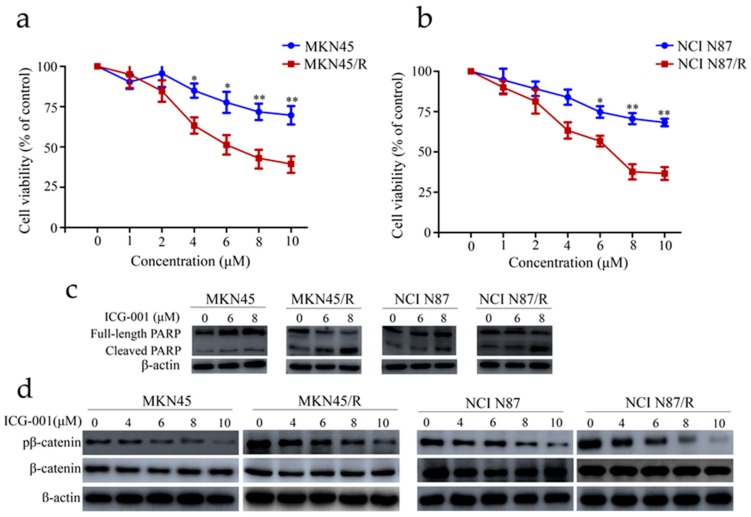
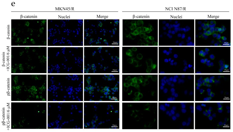
Figure 5.
ICG-001 preferentially inhibited cell viability and phosphorylation of β-catenin and induced apoptosis in trastuzumab resistant cells. (a,b) Cells cultured for 24 h were either treated with or without ICG-001 at the indicated concentrations or (dimethyl sulfoxide) DMSO, which was set as control. Cell viability was determined by CCK-8 assay 72 h later. Results expressed as % control represented the mean of three experiments. * p < 0.05, ** p < 0.01 vs. controls; (c) Cells exposed to ICG-001 for 24 h under different concentrations of (0, 6, 8 μM) were analyzed by Western blot for full-length PARP and cleaved PARP, with β-actin being a loading control; (d) Cells were exposed to ICG-001 for 24 h at the indicated concentrations and with DMSO as control, and cell lysates were probed with phosphor- and total antibodies of β-catenin signaling pathway. β-actin was used as a loading control. The representative images were cropped and shown; (e) MKN45/R and NCI N87/R cells were treated with ICG-001 (6 μM) for 24 h and DMSO as control which was followed by collection for immunofluorescence. Nuclear staining was performed using DAPI (blue), and total β-catenin and phosphor β-catenin were stained by Alexa Fluor® 488-labeled secondary antibody (green). Slides were imaged using ×20 lens of Leica DMi8 fluorescent microscope.


