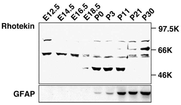
Figure 3.
Developmental change of Rhotekin in the rat brain. Cholate extracts of brains at various stages were subjected to SDS-PAGE followed by Western blotting with anti-Rhotekin (top) or anti-GFAP (bottom). Molecular markers are at right. (Adapted from Reference [7]).

