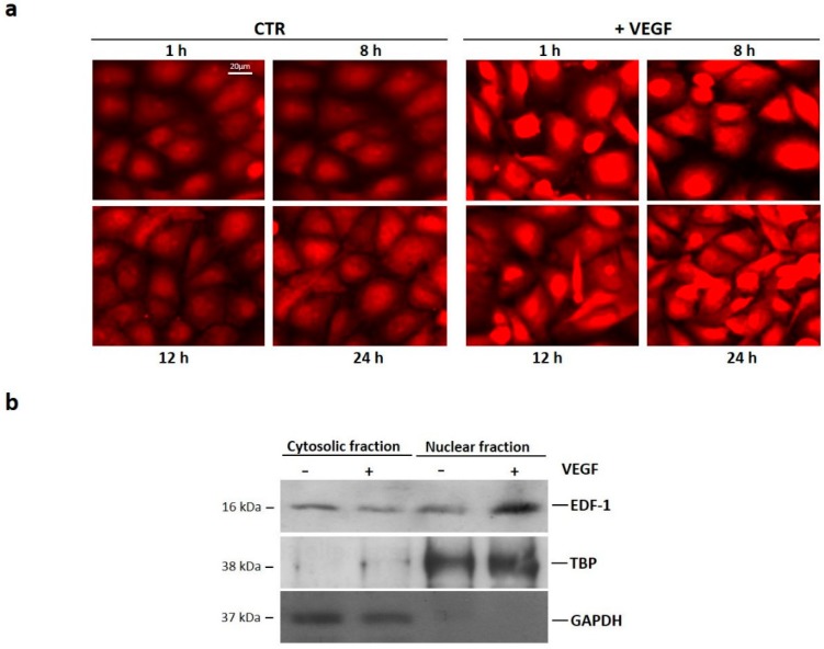
Figure 2.
Subcellular localization of EDF1 in cells treated with VEGF. (a) HUVEC were treated with VEGF (50 ng/mL) for 1, 8, 12, and 24 h. Immunofluorescence was performed using anti-EDF1 immunopurified immunoglobulin G (IgGs) and rhodamine-conjugated anti-rabbit IgGs; (b) HUVEC were treated with VEGF (50 ng/mL) for 1 h. Western blot was performed on nuclear and cytosolic fractions using antibodies against EDF1. GAPDH and TBP were used as cytosolic and nuclear markers, respectively. A representative blot is shown.

