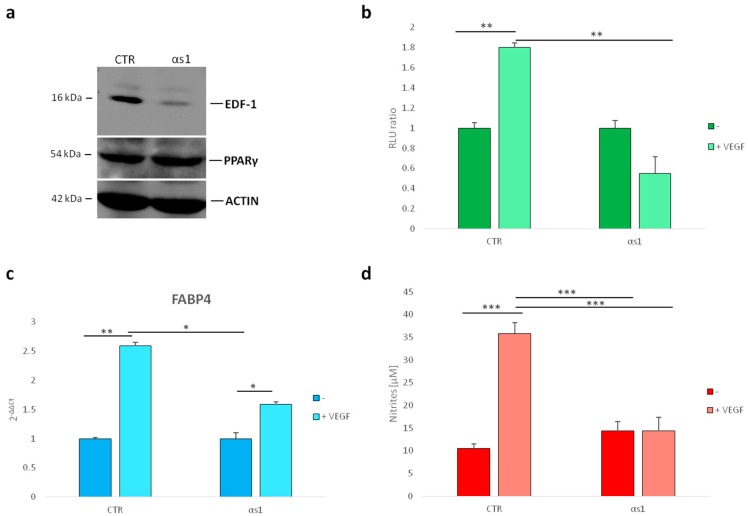
Figure 4.
PPARγ transcriptional activity in HUVEC with silenced EDF1. (a) The modulation of PPARγ was evaluated in αs1 cells (αs1) (HUVEC with stably silenced EDF1) and compared to HUVEC transfected with a scrambled nonsilencing sequence (used as control) (CTR). Cell lysates were analyzed by western blot using antibodies against EDF1, PPARγ, and actin. A representative blot is shown; (b) PPARγ activity was evaluated by luciferase assay in αs1 cells and compared to the control HUVEC; (c) Real-Time PCR was performed on RNA samples from αs1 cells and relative control, treated or not with VEGF (50/ng/mL) for 24 h. Three different experiments in triplicate were performed; (d) Nitric oxide (NO) release was measured using the Griess method for nitrate quantification. The values were expressed as the mean of three different experiments in triplicate ± standard deviation. * p < 0.05, ** p < 0.01, *** p < 0.001.

