Figure 1.
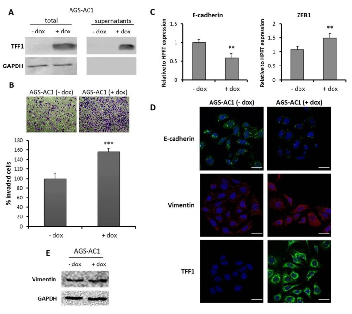
Trefoil factor 1 (TFF1) promotes invasion and epithelial to mesenchymal transition (EMT) changes in cellular models. (A) Protein level of TFF1 detected by western blotting. Protein normalization was performed on GAPDH levels; (B) Trans-well invasion assay of AGS-AC1 (TFF1 inducible hyperexpressing clone). Upper panel, bottom surface of filters stained with crystal violet. Magnification 10×. Bar = 100 μm. Lower panel, quantification of cell invasion. Statistically significant differences at p < 0.001 from the controls are indicated (***); (C) qRT-PCR for E-cadherin and ZEB1 mRNA expression in AGS-AC1 cells normalized on HPRT mRNA levels. Statistically significant differences at p < 0.01 from the non-induced cells are indicated (**); (D) Immunofluorescence analysis of TFF1 and vimentin on AGS-AC1 cells +/− doxycycline (induced or not induced to hyperexpress TFF1). Immunofluorescence images refer to 48 h after induction. Nuclei were stained with DAPI. Magnification 63×. Bar = 10 μm; (E) western blot analysis of vimentin expression in AGS-AC1 cells +/− doxycycline. Protein bands were normalized on GAPDH levels.

