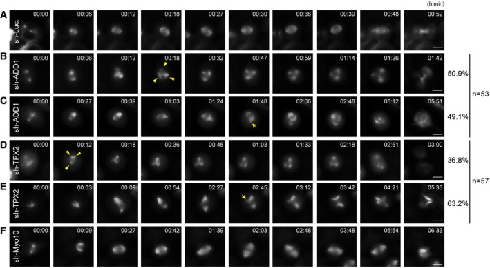
Figure EV4. Mitotic arrest contributes to, but does not fully explain ADD1 depletion‐induced spindle multipolarity.

-
A–FHeLa cells stably expressing mCherry‐tubulin were infected with lentiviruses expressing shRNAs to (A) luciferase (sh‐Luc.) as a control, (B, C) ADD1 (sh‐ADD1), (D, E) TPX2 (sh‐TPX2), or (F) Myo10 (sh‐Myo10). The cells were monitored with time‐lapse microscopy. Images were captured every 3 min for 18 h. Arrowheads indicate the multiple spindle poles occurs within 1 h upon nuclear envelope breakdown. Arrows indicate the multiple spindle poles occurs more than 1 h after entering mitosis. Scale bars, 10 μm. The percentages of these two categories in the total counted samples (53 for sh‐ADD1 and 57 for sh‐TPX2) are indicated to the right of the micrographs.
