
Figure EV1. Characterisation of SILAC‐labelled ES cells. Related to Fig 1 .
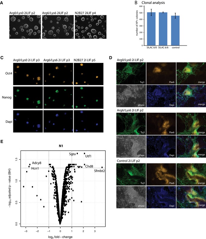
Cells were grown in 2iLIF for two passages before transfer into light (Arg0/Lys0) and heavy (Arg6/Lys6) SILAC medium.
- Phase contrast images of self‐renewing ES cell colonies in defined SILAC medium supplemented with 2iLIF. Arg0 = normal arginine; Lys0 = normal lysine; Arg6 = heavy arginine containing 6 × 13C, Lys6 = heavy lysine containing 6 × 13C. 20× magnification.
- Histogram showing number of alkaline phosphatase‐positive colonies formed from single cells deposited in 2iLIF after labelling for 2 passages in light (SILAC 0/0), heavy (SILAC 6/6) or full N2B27 (without Neurobasal)/2iLIF (control) medium. Mean and SD shown; n = 2.
- Immunostaining of SILAC‐labelled cells with Oct4 and Nanog antibodies after three passages in SILAC medium. 20× magnification.
- Immunostaining of SILAC‐labelled ES cells with Pax6 and Tuj1 antibodies on day 9 of culture in N2B27. Note: Arg6/Lys6 cells were treated with Chiron and LIF for 24 h before clonal analysis and gene expression profiling. p, passage. 20× magnification.
- Volcano blot illustrating fold changes and statistical significance for identified phosphorylated peptides in the nuclei fraction (N1). Results are from protein identifications in three independent eperiments.