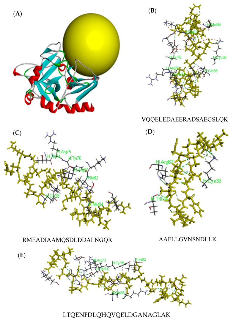
Figure 3.
Molecular docking for the interactions of peptide against Thrombin. Molecular structure of thrombin from PDB database (A), the yellow sphere showed the docking position; VQQELEDAEERADSAEGSLQK (B), RMEADIAAMQSDLDDALNGQR (C), AAFLLGVNSNDLLK (D), and LTQENFDLQHQVQELDGANAGLAK (E). The peptides were marked with yellow sticks; the other sticks are the interactive amino acids of thrombin.

