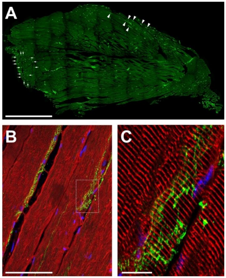
Figure 1.
Sympathetic innervation is richly developed in mouse hindleg muscle. Tibialis anterior muscles of adult wildtype mice were cryosectioned longitudinally, then stained with antibodies against TH (A–C, green) and, in addition, against α-actinin (B,C, red). Nuclei were stained with 4′,6-diamidino-2-phenylindole (DAPI; B,C, blue). Pictures show single confocal sections, (C) depicts the detail in the boxed region of (B). Arrowheads and arrows in (A) indicate alignment of sympathetic neurons with longitudinally and vertically cut muscle fibers, respectively. Scalebars show 500 µm, 50 µm, and 5 µm in (A–C), respectively.

