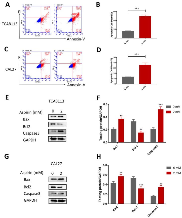
Figure 4.
Aspirin promoted apoptosis in the TCA8113 and CAL27 cells. (A,B) The apoptosis cell rates were increased with ASA treatment in the TCA8113 cells. The apoptotic cells were detected by flow cytometry after staining with Annexin V and PI after cells were treated with 2 mM ASA for 24 h. (C,D) Similar results were observed in CAL27. (E,F) The ASA treatment increased the expression of Bax and caspase3, and inhibited the expression of Bcl-2. Apoptosis related protein levels were detected by Western blotting using indicated antibodies, and GAPDH was used as a loading control. (G,H) Similar results were observed in CAL27. ** p < 0.01 and *** p < 0.005. The data were presented as the mean ± SD (n = 3).

