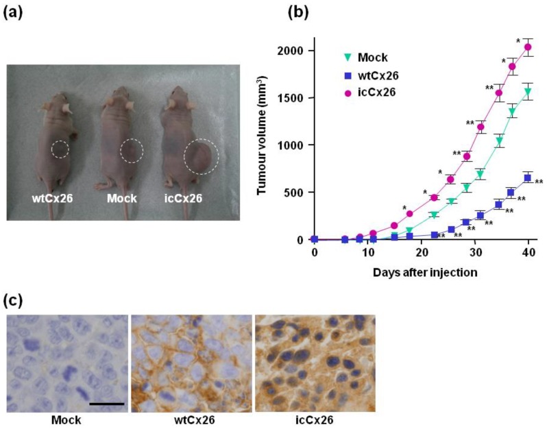
Figure 4.
Xenografts of the wtCx26, icCx26, and mock clones into nude mice and tumorigenicity assay in vivo. (a) Representative mice bearing tumours raised from 1 × 10⁶ cells of each clone. (b) Tumorigenicity in vivo of each clone. The size of each tumour was measured every 2 or 3 days. Error bars represent the SD (n = 6). No error bar is indicated when the SD is too small to show. * p < 0.03, ** p < 0.001 (significantly different from the mock clone at the corresponding time point). (c) Expression and subcellular localisation of Cx26 protein in the tumours raised from the xenografts. As revealed by immnohistochemistry, wtCx26 was localised in a cell-cell boundary area. Scale bar, 20 µm.

