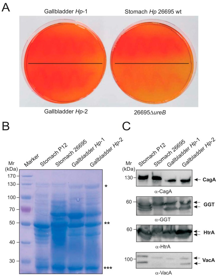
Figure 3.
Urease test and Western blotting analysis of H. pylori-specific pathogenicity factors from gallbladder strains. (A) Two H. pylori isolates (Gallbladder Hp-1 and Hp-2) were grown on acidified agar supplemented with urea (left samples). The observed color change from orange to red indicated that bacterial colonies were producing functional urease. The right samples represent positive controls. The color change occurred with the wild-type (wt) strain 26695 as expected, and was not observed with the negative control of an isogenic ΔureB deletion mutant, indicating that functional urease enzyme was not being produced; (B) Protein profiling using Coomassie staining. Asterisks label the following protein bands: CagA (*), Urease B (**), and Urease A (***); (C) Western blots of two reference strains (P12 and 26695) and the two gallbladder isolates that identifies presence of H. pylori proteins CagA, VacA, GGT and HtrA.

