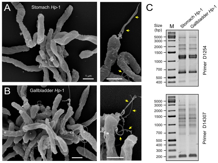
Figure 4.
Scanning electron microscopic and genetic analyses of the stomach and gallbladder H. pylori isolates. High resolution scanning electron microscopy of the cultures obtained from the stomach (A) and gallbladder (B) of the same patient revealed spiral-shaped H. pylori bacteria. Arrows in the enlarged sections indicate typical monopolar flagella being present; (C) PCR-based randomly amplified polymorphic DNA (RAPD) produced identical fingerprints for the two H. pylori strains isolated from stomach and gallbladder. This method uses a set of single indicated primers (D1254 or D14307, top and bottom), which arbitrarily anneal and amplify genomic DNA resulting in strain-specific fingerprinting patterns. M = DNA size marker.

