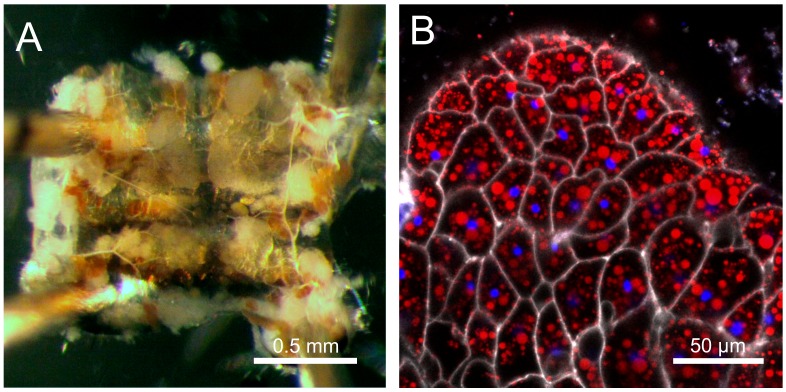
Figure 2.
The fat body of Drosophila. (A) Bright-field image of a subcuticular fat body (white tissue) attached to the abdominal cuticle. (B) Confocal microscope image of the fat body. Cell membranes in white (CellMask Deep Red), lipid droplets in red (BODIPY 493/503) and DNA in blue (Hoechst 33342).

