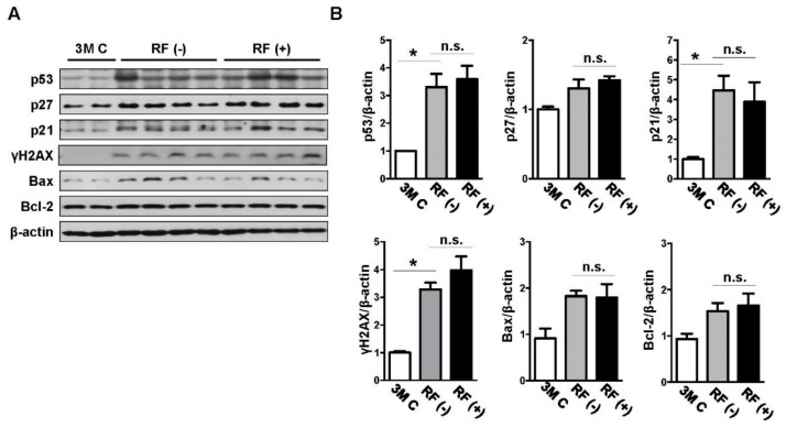
Figure 3.
Effects of RF-EMF exposure on the protein expression levels of p53, p27, p21, γH2AX, Bax, and Bcl-2 as determined by western blotting analysis. (A) Western blot images of DNA damage-related proteins in the mouse brain following RF-EMF exposure. (B) The graphs show the quantification of p53, p27, p21, γH2AX, Bax, and Bcl-2 protein levels in young-aged (3M C), sham-exposed (RF (−)), and RF-exposed (RF (+)) mouse brains based on band intensity. The values are presented as means ± SEM. * p < 0.05 versus young-aged (3M C) group. n.s.: no significance.

