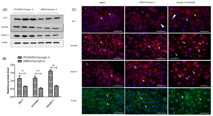
Figure 6.
The inhibitory effects of MEK/ERK on TJ protein expression, membrane distribution and F-actin polymerization. Western blotting analysis of ZO-1, occludin and claudin-1 expression ((A,B), n = 3); cell immunofluorescence ((C), n = 3, 400×) showed the membrane distribution (a–i) and F-actin polymerization (j–l). The results were confirmed by three independent experiments per treatment. Representative results of the three independent experiments are shown. Data (mean ± SEM) were analyzed with the Student’s t-test. Cell nuclei were stained by DAPI and are shown in blue. Claudin-1, ZO-1 and occludin are shown in red and pointed out by white arrow heads. F-actin is shown in green. Scale bar is 50 μm. ** p < 0.01, *** p < 0.001.

