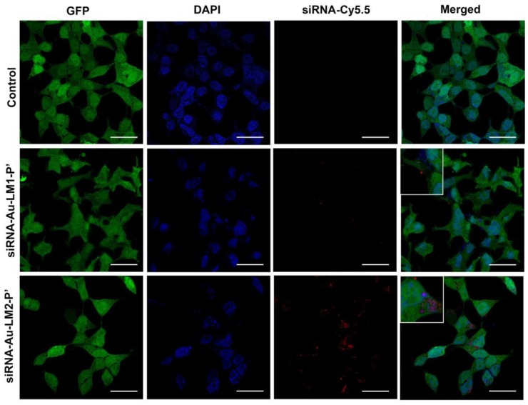
Figure 10.
Uptake of siRNA-Au-LM1-P’ and siRNA-Au-LM2-P’ nanoconstructions by HEK Phoenix cells. siRNA was labeled with Cy5.5 and observed as red spots. Nuclei were stained blue with DAPI and green GFP was diffusely located inside whole cells. In the merged image, location of siRNA in cytoplasm was shown (red spots). Length of scale bars correspond to 20 µm.

