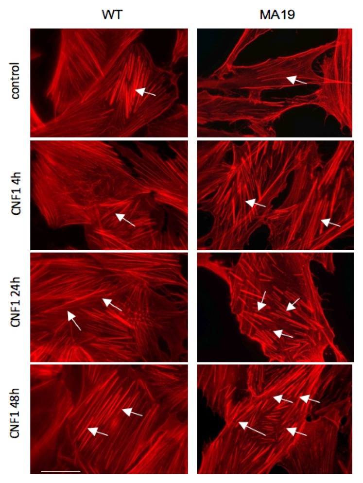
Figure 2.
In the MA19 patient’s fibroblasts, CNF1 restores the actin stress fibers’ organization typical of WT cells. Fluorescence micrographs of WT fibroblasts (left) column and MA19 fibroblasts (right) column treated with CNF1 at the indicated time points. Note that the actin stress fibers’ organization appeared improved in WT fibroblasts with an apparent reinforcement of the actin fibers. The reorganization promoted by the toxin was more evident in pathological fibroblasts that showed an impressive restoration of the cytoskeletal phenotype typical of fibroblasts. Arrows show the stress fibers. Bar = 10 µm.

