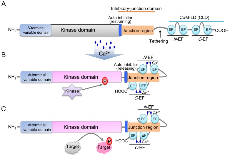
Figure 2.
Diagram of CDPK structure and the Ca2+-CDPK decoding Mechanism (modified from [68]). (A) Inactive state of the CDPK protein. The auto-inhibitor restrains the kinase activity, while EF-hand motifs do not bind to the junction region. (B) Active state of the CDPK protein. After Ca2+ uploading into elongation factor (EF) hands, N-EF and C-EF hands combine with different sides of junction region, then the kinase domain is released and phosphorylated simultaneously by an upstream kinase. (C) Phosphorylation of target proteins.

