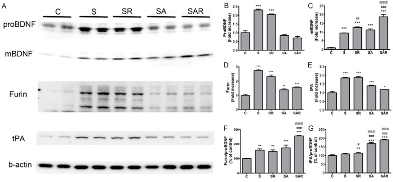
Figure 7.
Effect of TST and TST combined with 5-Aza-dC treatment on the expression of mature BDNF in protein extracts of contralesional cortex after stroke. (A) Representative photomicrographs of western blots for pro-brain-derived neurotrophic factor (proBDNF), mature BDNF (mBDNF), furin, tissue plasminogen activator (tPA), and β-actin in total lysates of the contralesional cortex at 4 w after post-stroke treatment. (B–E) Signal intensities of proBDNF, mBDNF, furin and tPA were measured using Quantity One software and are shown as a percentage of control. (F,G) Signal intensities of the ratio of furin to proBDNF and tPA to proBDNF are shown as a percentage of control. Beta-actin, internal control. Results are presented as the mean ± SE, n = 6. * p < 0.05; ** p < 0.01; *** p < 0.001 vs. C, # p < 0.05; ## p < 0.01; ### p < 0.001 vs. S, @@@ p < 0.001 vs. SR.

