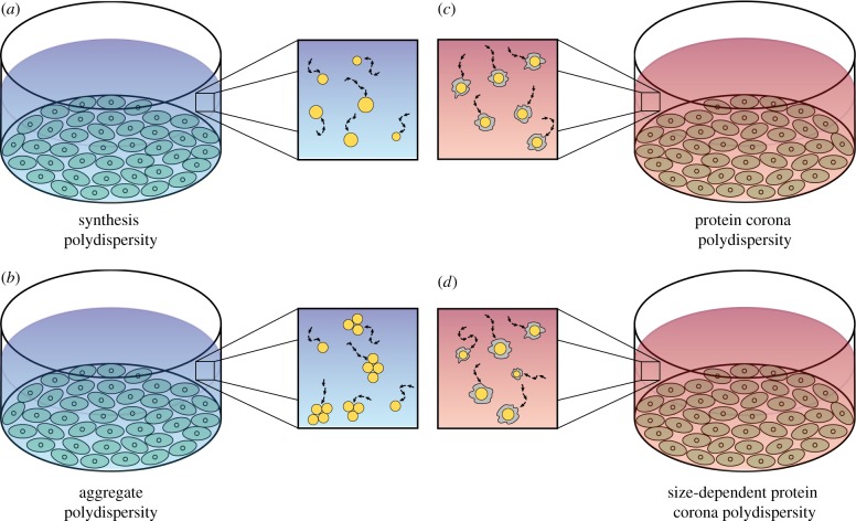
Figure 2.
(a–d) Schematic of the four types of nanoparticle polydispersity considered, in the context of an in vitro cellular association experiment. Blue backgrounds denote protein-free media, whereas red backgrounds denote protein-rich media. Orange circles represent nanoparticles and grey shapes represent the protein corona. (Online version in colour.)

