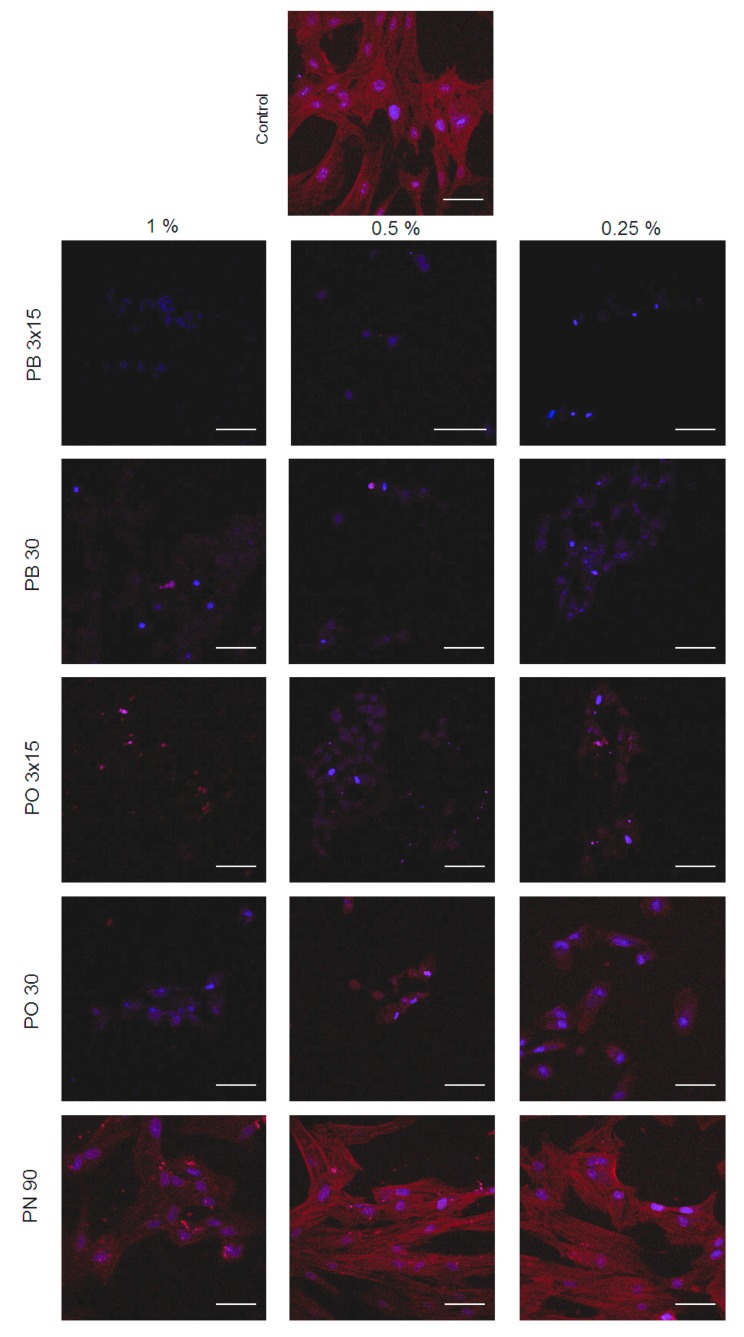
Figure 5.
Representative immunofluorescence micrographs revealing the cytoskeletal organisation of hDPSCs exposed to control and bleaching products. Cultured hDPSCs were treated with undiluted extracts of bleaching products for 24 h. For staining filamentous actin (F-actin), cells were incubated with CruzFluor594-conjugated phalloidin (Santa Cruz Biotechnology, Dallas, TX, USA). Nuclei were counterstained with 4,6-diamidino-2-phenylindole dihydrochloride (DAPI) (blue). Scale bar = 150 μm.

