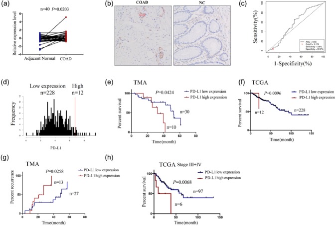
Figure 1.
Increasing expression of PD-L1 predicts poor survival and recurrence in patients with COAD. (a) TMA analysis of the differentially expressed level of PD-L1 in pair-matched tumor tissues. (b) IHC analysis of the expression level and cellular localization of PD-L1 in COAD tissue cells (200×). (c) The cut-off value and AUC of PD-L1 were assessed in COAD samples (n = 240). (d) The patients with COAD were divided into PD-L1 high expression group and low expression group. (e) Kaplan–Meier analysis of the link of PD-L1 high or low expression with the OS in patients with COAD from TMA data. (f) Kaplan–Meier analysis of the link of PD-L1 high or low expression with the OS in patients with COAD from TCGA data. (g) Kaplan–Meier analysis of the link of PD-L1 high or low expression with the tumor recurrence in patients with COAD from TMA data. (h) Kaplan–Meier plotter analysis of the correlation of PD-L1 high or low expression with the OS of COAD patients with stage III + IV.

