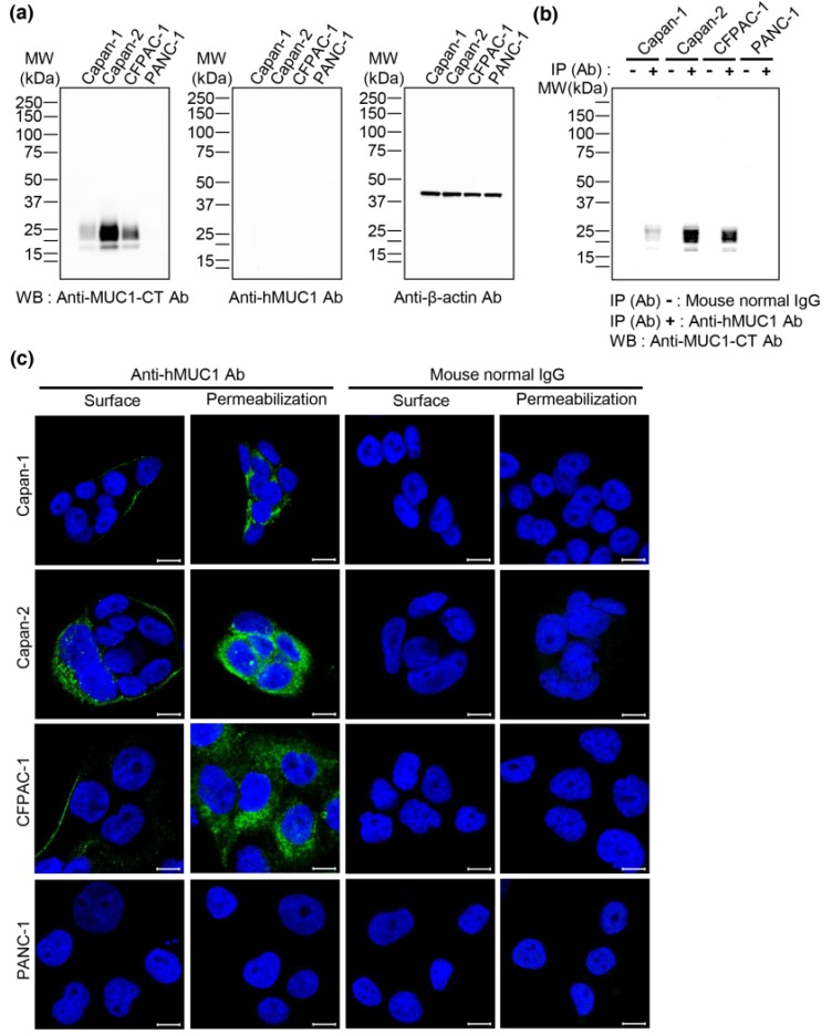
Figure 1.
Immunoreactivity of anti-hMUC1 monoclonal antibody in pancreatic cancer cells. (a) Lysates from the pancreatic cancer cell lines Capan-1, Capan-2, CFPAC-1 and PANC-1 were analyzed by western blotting using anti-MUC1-CT, anti-hMUC1 and anti-β-actin antibodies. (b) Capan-1, Capan-2, CFPAC-1 and PANC-1 cell lysates were immunoprecipitated with mouse normal IgG or anti-hMUC1 monoclonal antibody followed by western blotting using anti-MUC1-CT antibody. (c) Capan-1, Capan-2, CFPAC-1 and PANC-1 cells were fixed with 4% paraformaldehyde and incubated with mouse normal IgG or anti-hMUC1 monoclonal antibody on ice to identify MUC1-C on the membrane surface of the cells (surface). On the other hand, to identify MUC1-C in the intracellular region, the cells were fixed, permeabilized and incubated with mouse normal IgG or anti-hMUC1 monoclonal antibody at room temperature (permeabilization). Then, the cells were stained with Alexa 488-conjugated secondary antibody. Hoechst 33258 was used for staining nuclei. Images were captured using confocal microscopy. Scale bars, 10 µm. These results are representatives of three independent experiments.

