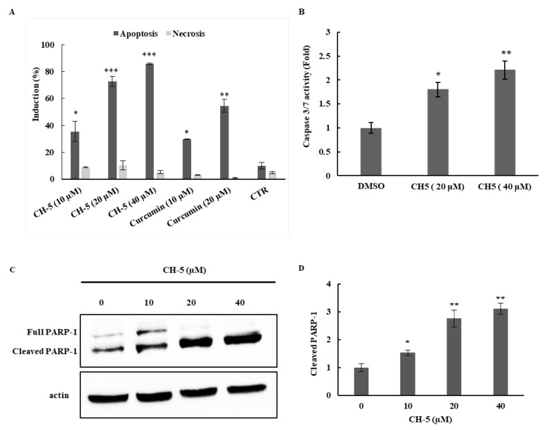
Figure 2.
(A) Analysis of apoptosis and necrosis induction in U2OS cells treated with CH-5 and curcumin at the indicated concentrations for 24 h. The cells were double-stained with Annexin–FITC and PI to identify apoptotic and necrotic cells; (B) Apoptosis was further confirmed by measuring caspase 3/7 activity in U2OS cells treated with CH-5. After treatment with CH-5 at the indicated concentrations, the cells were collected and supplied with the substrate solution for caspase-3/7 activity determination, according to the manufacturer’s instructions, as described in materials and methods; (C) Western blot analysis of PARP-1 cleavage in U2OS cells treated with CH-5. U2OS were cultured and treated as described in 1A. Western blots of intact PARP-1 and cleaved PARP-1 in CH-5-treated and control (DMSO)-treated cells are shown; (D) The intensity of the PARP-1 fragment due to caspase activation was dose-dependent. The image intensities of the bands were normalized against the intensity of the β-actin band. The data are expressed as mean values ± SD (n = 3); * p < 0.05, ** p < 0.01 and *** p < 0.001 indicate a significant difference with respect to the control.

