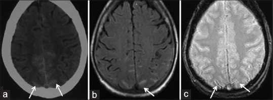
Figure 3.

Axial sections of the brain reveal hyperdensity (a, computed tomography), hyperintensity on fluid-attenuated inversion recovery (b, MRI) along parafalcine sulci. (c) depicts corresponding susceptibility-weighted image. These findings confirm the presence of subarachnoid hemorrhage
