Figure 1.
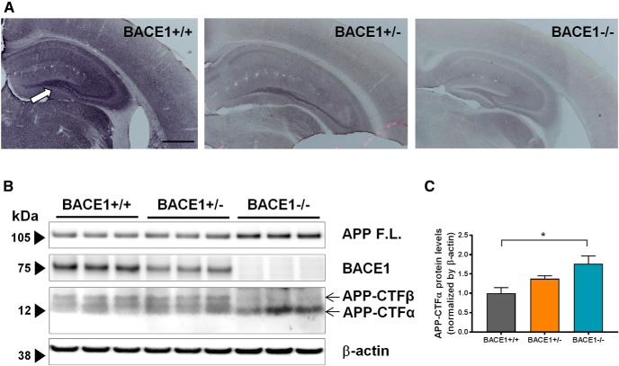
BACE1 is not expressed in BACE1–/– mice. A, BACE1 expression was assessed in coronal sections of 2-month-old BACE1+/+ (left), BACE1+/– (middle), and BACE1–/– (right) mice using immunohistochemistry with a commercial BACE1 antibody. BACE1 immunoreactivity is present throughout BACE1+/+ sections, with higher levels in the mossy fiber pathway of the hippocampus (white arrow). Scale bar, 500 µm. Conversely, there is no BACE1 immunoreactivity in BACE1–/– brain sections. B, BACE1 expression is attenuated in BACE1+/– and abrogated in BACE1–/– mice. The expression and activity of BACE1 was assessed in BACE1+/+, BACE1+/–, and BACE1–/– mice with Western blot. BACE1 is expressed robustly in BACE1+/+ brain tissues, is weakly expressed in BACE1+/– brain tissues, and is not expressed in BACE1–/– brain tissues, while β-actin controls are expressed equally in all three. APP-CTFα, a product of the enzymatic cleaving of APP by α secretase, is present in tissues from all three genotypes. However, APP-CTFβ, a product of the enzymatic cleaving of APP by BACE1, is present in BACE1+/+ and BACE1+/– tissues, but not in BACE1–/– tissues. C, Densitometric analysis of APP-CTFα. F(2,6) = 6.557, p = 0.0309; *, p < 0.05.

