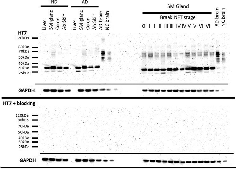
Figure 2.

Tau Western blots of peripheral organs (left) and submandibular gland tissues from a variety of Braak NFT stages (right) of 30ug of tissue and 1ug of brain probed with a HT7 (top) and probed with HT7 and a blocking peptide (10x the concentration of HT7) directed towards the HT7 sequence. Boxes below each respective blot represent GAPDH of that blot.
