
Figure 4.
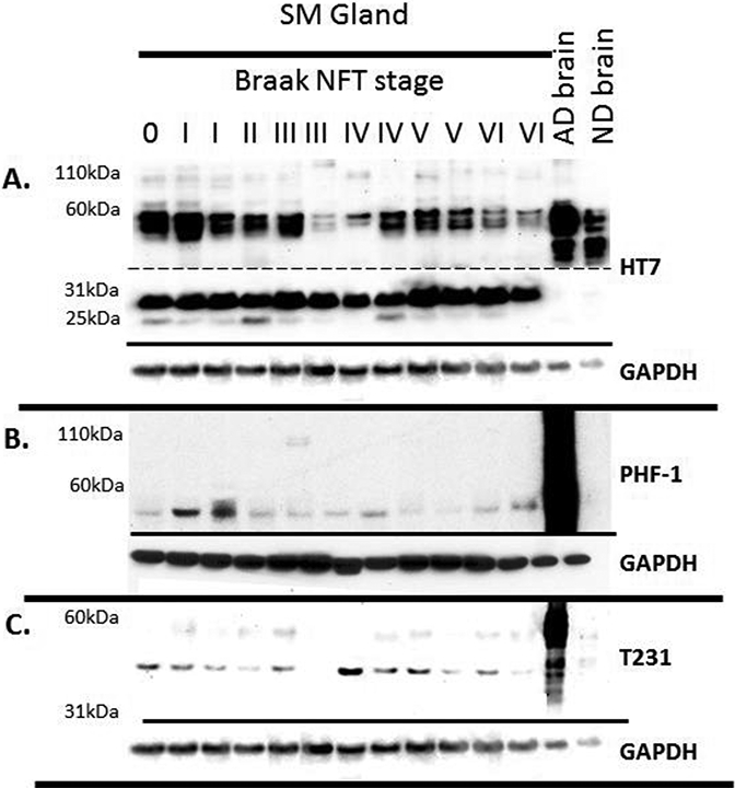
Tau Western blots of submandibular gland tissues from a variety of Braak NFT stages. 30ug of submandibular gland and 1ug of brain were run on 4–12% Bis-Tris gels and probed with A) HT7 (dashed line indicates different exposure times: Top long exposure, bottom short exposure), B) PHF-1, C) and T231. GAPDH was used as a protein loading control and is located below each blot.