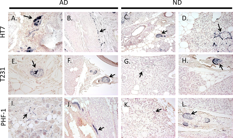
Figure 7.

Examples of immunoreactive HT7 (A-D), T231 (E-H), and PHF-1 (I-L) nerve elements (black arrows) within submandibular glands of AD (left) and ND (right) cases. Immunoreactive ganglion cells (A, E), nerve fibers surrounding an arteriole (B), stromal nerve fascicles (A, C, E, F, H, J, L), and nerve fibers intertwined amongst glandular acini (D, G, I, K). Photos were taken at 40x magnification, except for C, F, and J which were taken at 20x.
