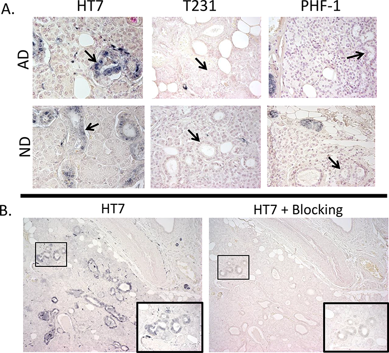
Figure 8.

A) Examples of simple cuboidal epithelial cell (black arrows) immunoreactivity with HT7, but not with PHF-1 or T231 within the submandibular gland of AD and ND cases; photos taken at 40x. B) Submandibular gland tissues stained with HT7 (left) and probed with HT7 and a blocking peptide (10x the concentration of HT7) directed towards the HT7 sequence (right) demonstrated diminished immunoreactivity; photos taken at 10X magnification. Images in insets show the corresponding boxed area at higher magnification.
