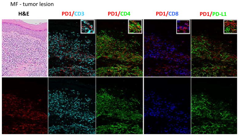
Figure 5. Histologic and immunophenotypic features of CTCL tumor, with 6-color multiplex immunohistochemistry for immune checkpoint expression.
Multiplex images performed on skin sections of MF lesion from a representative patient with tumor lesion (T3) is shown. PD-1 co-localizes with CD3, CD4, and CD8 expression. PD-1 and PD-L1 expression do not overlap. Higher expression of PD-1 and PD-L1 expression is noted on tumor lesion (T3), compared with plaque (T2, Fig. 4). [200x magnification; insets represent 400x magnification of small regions of multiplex images]. Representative data from 3 independent experiments are shown.

