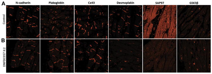
FIGURE 5. Immunohistochemistry of Left Ventricular Myocardial Tissue.
Immunostaining of the explanted heart of patient DNFDC057-II:2 carrying the G1891Vfs61X FLNC truncation. (A) Immunoreactive signals for plakoglobin and connexin 43 (Cx43) at intercalated discs are normal compared with control samples, whereas junctional signal for desmoplakin is reduced. N-cadherin is used as a tissue quality control and is normal in all samples. (B) SAP97 signal is depressed compared with control samples. GSK3β maintained its normal cytoplasmic distribution.

