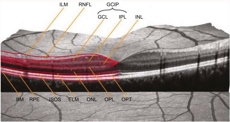
Figure 1.
Macular OCT with intraretinal layers.
Notes: Reproduced from Schematic Figure – Macular OCT with Intraretinal Layers by Neurodiagnostics Laboratory @ Charité – Universitätsmedizin Berlin, Germany. Available from: http://neurodial.de/2017/08/25/schematic-figure-macular-oct-with-intraretinal-layers/. Creative Commons Attribution 4.0 International License.65
Abbreviations: OCT, optical coherence tomography; ILM, internal limiting membrane; RNFL, retinal nerve fiber layer; GCIP, ganglion cell–internal plexiform; GCL, ganglion cell layer; IPL, internal plexiform layer; INL, inner nuclear layer; BM, Bruch membrane; RPE, retinal pigment epithelium; ISOS, inner segment–outer segment (junction); ELM, external limiting membrane; ONL, outer nuclear layer; OPL, outer plexiform layer; OPT, outer photoreceptor tip.

