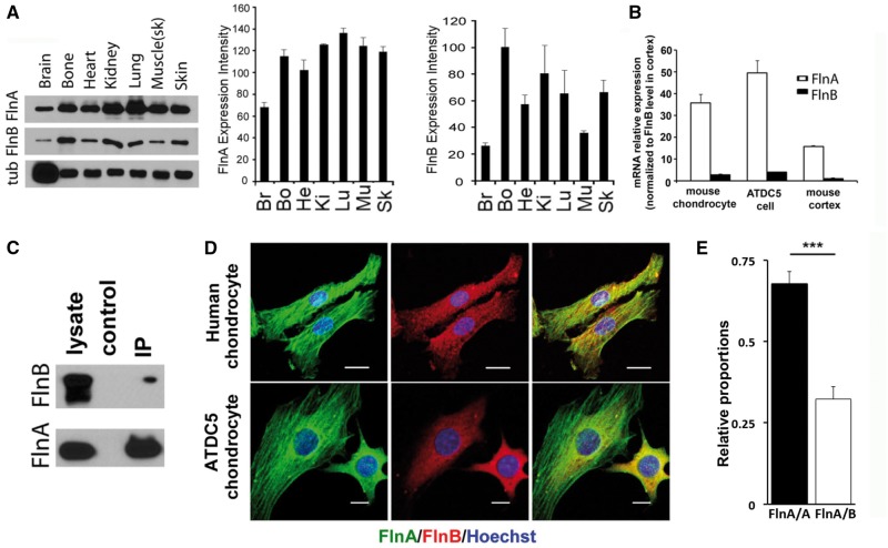
Figure 1.
FlnA and FlnB expression pattern, interaction and co-localization. (A) Western blot analyses shows widespread endogenous expression levels for both FlnA in multiple tissues of postnatal day 1 wild type mice. FlnB protein shows relatively higher expression levels in bone and kidneys. (B) Quantitative RT-PCR shows that mRNA expression for FlnA is higher in primary mouse chondrocytes, ATDC5 chondrocytes, and murine cerebral cortex compared to FlnB. (C) Endogenous FlnA immunoprecipitation by FlnB in mouse ATDC5 cells indicate that these two proteins interact in proliferating chondrocytes. Mouse anti-FlnA antibody and rabbit anti-FlnB were used to detect the input and immunoprecipitated filamin proteins. The specificity for these antibodies has been previously demonstrated in the null FlnA/B mice (10,36). (D) Fluorescent photomicrographs show overlapping expression for both FlnA and FlnB in the cell cytoplasm for primary human and mouse ATDC5 chondrocytes. FlnA also localizes to the actin stress fibers. Scale bars = 20 μm for human chondrocytes and 10 μm for mouse ATDC5 cells in D. (E) FlnA/A homo-dimers and FlnA/B heterodimers exist at an approximate ratio of 2:1 in HEK293 cells. *P≤0.05,**P < 0.01, ***P <0.001.

